Empower Your Application With CryoViz Imaging Service
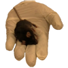
CryoViz imaging now can provide you high-detail answers over mouse-sized volumes! In 3D. In real color. With microscopic resolution.You can avail this novel 3D technology through a unique fee-for-service model. In summary, CryoViz imaging provides you the following benefits:
MICROSCOPIC COLOR ANATOMY
No more low resolution gray-scale images with limited contrast
SINGLE CELL DETECTION
Identify even single stem and cancer cells anywhere in a mouse
MULTI-SPECTRAL FLUORESCENCE
Label target cells in one color and imaging agent with another color to study targeting
MULTI-SCALE VISUALIZATION
From whole mouse to organs to tissues to cells—query your specimen in the digital domain at any resolution
You Ship Us Samples. We Deliver Results. Simple
 |
Design Your Experiment |
 |
 |
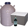
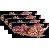
Prepare Sample |
 |
 |
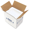
Ship To Us |
 |
 |
We Image & Analyze |
 |
 |
Deliver Results |
 |
 |
You Infer and Conclude |
 |